42 labels of the human brain
Amazon.com: brain model labeled VEVOR Human Brain Model Anatomy 4-Part Model of Brain w/Labels & Display Base Color-Coded Life Size Human Brain Anatomical Model Brain Teaching Human Brain for Science Classroom Study Display Model 3 $159 19 Get it Wed, Mar 30 - Mon, Apr 4 FREE Shipping Human Brain Model for Neuroscience Teaching with Labels 2 ... Human Brain Model for Neuroscience Teaching with Labels 2 Times Life Size Anatomy Model for Learning Science Classroom Study Display Medical Model: ...Theme: Anatomy,ScienceManufacturer: BEAMNOVABrand: BEAMNOVASize: 35.5x28x20cm (14x11x8 in) Rating: 4,1 · 28 reviews
Glucose and The Brain: Improving Mental Performance | Eufic 30.04.2013 · The human brain is made up of a dense network of neurons, or nerve cells, which are constantly active — even during sleep. To obtain the energy needed to sustain this activity, the brain depends on a continuous supply of glucose from the bloodstream. A healthy diet should provide 45-60% of total energy from carbohydrates.

Labels of the human brain
A unified 3D map of microscopic architecture and MRI of the human brain 27.04.2022 · The inclusion of five microscopy labels, blockface images, and three quantitative MRI contrasts provides a wealth of anatomical information ().The full-brain coverage allows for detailed and comparative analyses of architectonic features for mapping the cortical laminar structure (20–23).A second important application is the atlasing of small brain structures that … › pmc › articlesMRI Segmentation of the Human Brain: Challenges, Methods, and ... Mar 01, 2015 · If an atlas or template of the human brain for a specific population of interest is available, then atlas-based methods can be a powerful tool for brain MRI segmentation. The atlas contains information about the brain anatomy (e.g., it contains the information about the location of different brain structures) and it is used as a reference (a ... 5 Lobes Of The Brain (A Complete Guide) - NeuroTray 5 lobes of the brain. Each cerebral hemisphere is divided into five lobes: the frontal lobe, the parietal lobe, the occipital lobe and the temporal lobe, four of which have the same name as the bone above them. Deep within the lateral sulcus lies a fifth lobe, the insula or Island of Reil.
Labels of the human brain. Labeled Human Brain Illustrations, Royalty-Free Vector ... Browse 96 labeled human brain stock illustrations and vector graphics available royalty-free, or start a new search to explore more great stock images and vector art. Newest results Brain functions vector illustration. Labeled explanation organ... Colored and labeled human brain diagram Diagram of a Brain detailed anatomy of the human brain. Brain Label - The Biology Corner Image of the brain showing its major features for students to practice labeling. Answers are included. Regionconnect: Rapidly extracting standardized brain ... Towards this goal, the present work first generated the white matter connectome of the IIT Human Brain Atlas v.5.0 (Zhang and Arfanakis, 2018), then introduced and developed multi-layer, connectivity-based labels for each white matter voxel of the atlas, and finally generated software that integrates the information in the multi-layer labels ... Brain (Human Anatomy): Picture, Function, Parts ... • The cortex is the outermost layer of brain cells. Thinking and voluntary movements begin in the cortex. • The brain stem is between the spinal cord and the rest of the brain. Basic functions like...
Solved Label the structures and lobes of the human brain ... Label the structures and lobes of the human brain by clicking and dragging the labels to the correct location. <--Anterior Posterior --> Precentral gyrus Temporal lobe Parieto-occipital sulcus Parietal lobe Lateral sulcus Insula Postcentral gyrus Central sulcus Occipital lobe Frontal lobe Reset Zoom Parts of the Human Brain | Anatomy & Function - Video ... The parts of the brain include the cerebrum, the cerebellum, the brain stem, and the pituitary gland. The brain structure is protected by the skull, which is composed of the cranium and the bones... Labeled Diagrams of the Human Brain You'll Want to Copy ... The central core consists of the thalamus, pons, cerebellum, reticular formation and medulla. These five regions are the central areas that regulate breathing, pulse, arousal, balance, sleep and early stages of processing sensory information. The thalamus interprets the sensory information and helps determine what is good and bad. Brain - Human Brain Diagrams and Detailed Information Stria Medullaris of Thalamus Thalamus (3rd Ventricle) Tuber Cinereum Cerebrum Angular Gyrus Anterior Commissure of Brain Calcarine Sulcus Central Sulcus of Rolando Cingulate Gyrus Cingulate Sulcus Corpus Callosum Cuneus Fornix of Brain Frontal Pole Inferior Frontal Gyrus Inferior Parietal Lobule Inferior Temporal Gyrus Lamina Terminalis
Human Brain Photos and Premium High Res ... - Getty Images Browse 27,513 human brain stock photos and images available, or search for human brain anatomy or human brain illustration to find more great stock photos and pictures. human brain icons - vector illustration - human brain stock illustrations. abstract brain activity image - human brain stock pictures, royalty-free photos & images. A single-cell atlas of the normal and malformed human brain … 27.01.2022 · Diagnosis of human brain AVMs were angiographically confirmed preoperatively, and fresh tissues were acquired as part of planned surgical resection. For scRNA-seq, only unruptured brain AVMs were enrolled into the study. Specimen orientation was maintained to ensure coverage of arteriovenous axis with surgical clips of different sizes with aide ... The Brain and Nervous System | Noba There are approximately 86 billion neurons in the human brain and each has many contacts with other neurons, ... [Image: Biology Corner, , CC-BY-NC-SA 2.0, , labels added] The cerebrum (also called the “cerebral cortex”) is the “newest,” most advanced portion of the brain. The cerebral hemispheres (the left and right … Human brain - Wikipedia The human brain is the central organ of the human nervous system, and with the spinal cord makes up the central nervous system.The brain consists of the cerebrum, the brainstem and the cerebellum.It controls most of the activities of the body, processing, integrating, and coordinating the information it receives from the sense organs, and making decisions as to the instructions sent to the ...
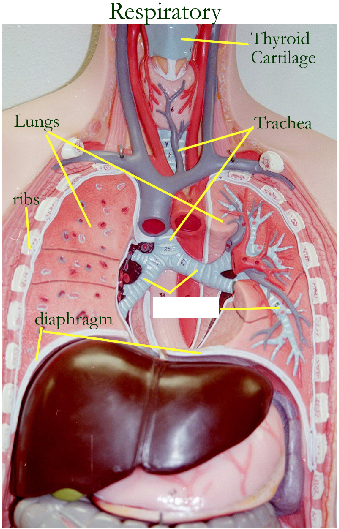
Print Activity 5: Examining the Human Torso Model flashcards | Easy Notecards
MRI Segmentation of the Human Brain: Challenges, Methods, … 01.03.2015 · If an atlas or template of the human brain for a specific population of interest is available, then atlas-based methods can be a powerful tool for brain MRI segmentation. The atlas contains information about the brain anatomy (e.g., it contains the information about the location of different brain structures) and it is used as a reference (a prior knowledge) for segmenting …

TDP-43 mutant transgenic mice develop features of ALS and frontotemporal lobar degeneration | PNAS
Labeled Brain Model Diagram | Science Trends The frontal lobe of the brain is responsible for our critical thinking, planning, reasoning, and problem-solving, as well as our experience of emotions. The rear portion of the frontal lobe is the motor cortex, which receives inputs from the other lobes and carries out the movements of the body associated with them.
Post a Comment for "42 labels of the human brain"